Education Center
Melasma in Skin of Color
Melasma is a condition that resulted from an increased accumulation of melanin. While it is an acquired condition, it is also one of the most common skin conditions that affect the dark-skinned population. The brown or light-colored skin population as in Asians and Latinos are among the most affected when considering people with skin of color. The statistics also show that melasma occurs more frequently in women than in men. This makes melasma a disease of women!
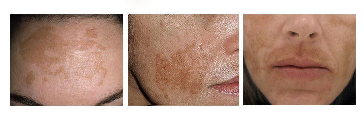
This disorder affects the areas of the body which are exposed to sunlight such as the face, neck, and shoulders. The areas involved are affected in three patterns: centrofacial pattern, malar pattern, and mandibular pattern. The centrofacial pattern includes the forehead, nose, upper lip, cheeks, and chin, while the malar pattern involves the area of the nose and cheeks. However, the mandibular pattern of the development of melasma includes only the sides of the lower jaw. Melasma usually follows only one of the above-stated three patterns of melanin accumulation (hyper melanosis).
Before going deeper into how melasma appears, it is better to know, why melasma develops or what causes this condition. The major causes of melasma are genetics, sunlight and ultraviolet radiation exposure, and hormones, especially during pregnancy. These trigger factors activate the increased production of melanin and as a result of it, hyper melanosis develops. This is the reason for melasma.
Melasma is usually tan or brown in color, but dermal melasma shows a black or blue color. The major element of melasma lesions is small brown flat skin patches. These macules are usually situated in the areas of the body where the greatest density of sebaceous glands is located.
Melasma is identified with the help of the clinical picture along with the help of a Wood’s lamp or skin scanner examination. These pieces of equipment help to differentiate between epidermal and dermal pigmentation in melasma, where the visibility of the melanin is only possible with an epidermal version of melasma. These data help to identify the condition accurately. The exact identification of melasma helps to initiate the treatment. The best treatment for this condition is the use of alpha hydroxy acid (AHA) and Alpha/Beta peels. This can be accompanied by the use of cosmetics for hyperpigmentation. Other chemical peels, serums, and laser treatments can also treat melasma. However, it is very important to know that even mild rays of sunlight have the power to induce this form of hyper melanosis. Therefore, sunscreens that are broad should be used as a prophylaxis. Apart from the mainstream treatment, vitamin A derivatives also show some improvement in controlling melasma. In addition, the other topical agents that can be used to decrease the level of pigment in melasma are Azelaic acid topical and Kojic acid topical, and ascorbic acid. There is some research that suggests that oral administration of proanthocyanidin along with vitamins and tranexamic acid are beneficial in decreasing the pigmentation in melasma, but they are prescription medications and only required if melasma is resistant to conservative therapy.
Because melasma has proven to be a challenge in its treatment, it is best a patient/client is seen weekly or bi-weekly. Home management products are important for clients to purchase. Using these products twice daily will maintain or enhance the in-clinic treatments. Optimal results will be seen in a shorter period of time. But, remember, to get optimal results, the patient/client must apply a skin lightening serum or cream to the affected areas every evening.